Medically reviewed by Dr. Prem Ratan Degawat, MD, DM (Cardiology)
Senior Interventional Cardiologist · Director, Mitral & Tricuspid Valve Therapy · Associate Director, TAVR & Structural Heart Disease Program, Eternal Hospital, Jaipur
Last updated on Nov 16, 2026 · View LinkedIn profile
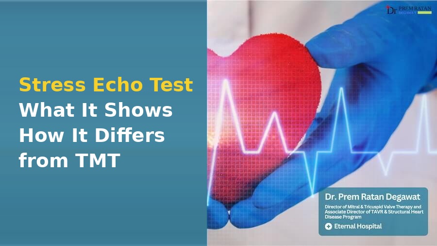
A stress echo test uses ultrasound pictures to compare how your heart pumps at rest and when it works harder. A standard TMT mainly records the heart’s electrical activity during exercise; a stress echo adds pictures of the moving heart muscle. It can help investigate certain symptoms or clarify another test, but not everyone with chest pain needs it.
What does a stress echo look for?
Your heart muscle needs more blood when it works harder. If a narrowed heart artery limits that supply, part of the muscle may squeeze less strongly during stress. The doctor compares pictures from different stages to look for that change. The medical term is inducible ischaemia, meaning signs of too little blood supply brought on by effort.
The test does not photograph every artery or give an exact percentage blockage. It asks whether the heart’s response suggests a blood supply problem. Your symptoms, exercise level and blood pressure also help the doctor understand the result.
The British Society of Echocardiography guideline describes this comparison of heart muscle movement as a central part of stress echo. Sometimes the test answers a different question, such as how a valve behaves under extra work.
How is it different from a normal echo and TMT?
A normal echo shows the heart while you are resting. It can show valve problems or reduced pumping strength, yet look normal when an artery only limits blood supply during exertion.
| Test | What it records | Main difference |
|---|---|---|
| Resting echo | Ultrasound pictures at rest | Shows structure, valves and resting pump function |
| Standard TMT | ECG during treadmill exercise | Looks at electrical changes and the response to exercise |
| Stress echo | Ultrasound pictures during or just after stress | Compares muscle movement at rest and with extra work |
ECG monitoring is also used during stress echo for safety. Adding pictures can help when an exercise ECG is unclear, but poor ultrasound views can limit the test too. The choice depends on the clinical question and which test you can complete safely.
Our TMT guide explains the treadmill ECG in more detail. The heart tests guide puts both tests alongside other investigations.
Who might be advised to have one?
A doctor may suggest stress echo for stable chest discomfort on exertion, selected cases of unexplained breathlessness, or symptoms in someone with known heart artery disease. It may help when an earlier test has left a question that would change treatment.
For example, a scan may show a narrowing without making clear whether it limits blood supply. Stress imaging can sometimes help answer that question. In other people, a CT scan of the heart arteries or another test may be more useful.
It is not a routine health check for everyone who feels well. Having risk factors does not automatically mean this is the right next test. Ask what your doctor hopes to learn and how the result could change your care.
Exercise or dobutamine stress echo?
For an exercise stress echo, you walk on a treadmill or pedal a special exercise bicycle. The effort rises in stages. With a treadmill, the team takes pictures quickly after you stop; with a suitable bicycle, pictures can be taken while you pedal.
If you cannot exercise enough, the team may use a medicine called dobutamine through a small tube in a vein. It makes the heart beat faster and more strongly while you lie on the examination couch. This is called a dobutamine stress echo.
Medicine stress is not an easier test that suits everybody. Your heartbeat, other medicines and overall condition affect the choice. Some people need a different investigation altogether.
How should you prepare?
Get instructions from the testing centre before the appointment. Food restrictions and medicine instructions differ with the test method and the reason for testing.
Bring your referral, earlier ECG and echo reports, and a list of everything you take. Tell the team about asthma, diabetes, drug allergies, pregnancy or a recent change in chest symptoms. For exercise testing, wear comfortable clothes and shoes suitable for walking.
Do not stop heart tablets on your own. Medicines that slow the heart can affect the test, but stopping them can also be unsafe. The BSE guideline says this decision needs to account for why the medicine was prescribed and what the test is meant to assess.
If you use insulin or other diabetes medicines and have been asked to avoid food, request a specific plan. Also ask how long to allow for the appointment and whether you need someone to accompany you home. Follow the centre’s advice for your test.
What happens in the test room?
The team checks your history, attaches ECG stickers and measures your blood pressure. An ultrasound probe with gel records the resting pictures. You may be asked to lie on your side or briefly hold your breath so the pictures are clear.
During exercise or the medicine infusion, staff watch your heartbeat, blood pressure and symptoms. Tell them promptly about chest discomfort, dizziness, unusual breathlessness or a racing heartbeat. You do not need to push through symptoms to produce a useful result.
The team stops when it has enough information or when symptoms, blood pressure or heartbeat changes make stopping necessary. Further pictures are taken during recovery. If the ultrasound view is poor, an injected contrast agent may help show the heart muscle more clearly. Mention any previous reaction to an injected agent.
Is a stress echo safe?
It is a supervised medical test with trained staff and emergency equipment available. Exercise or dobutamine can cause chest discomfort, blood pressure changes or an abnormal heartbeat. Serious complications are uncommon, but the test is not risk-free.
The team must reassess whether testing is appropriate if your symptoms have become unstable. Severe symptomatic aortic valve narrowing, uncontrolled heart failure or certain dangerous heartbeats may make a standard stress test unsafe.
Do not wait for a booked stress echo if you develop sudden severe chest pain, collapse or severe breathlessness. Seek emergency help, especially if pain spreads to your arm or jaw or comes with sweating. Do not drive yourself.
What do normal, abnormal and inconclusive results mean?
A normal result means no concerning stress-related change was found under the conditions achieved. That is reassuring, especially when the pictures were clear and the heart worked hard enough. It cannot rule out every kind of heart disease or explain every symptom.
An abnormal result may suggest that part of the heart receives too little blood during effort. The doctor considers where the change occurred, how much muscle was involved and your symptoms. An abnormal result does not automatically mean you need a stent.
An inconclusive test leaves the question open. Perhaps the pictures were unclear, the heart did not reach enough workload, or the test had to stop early. The report should explain the limitation. Your doctor may suggest another type of imaging or review the treatment plan.
Persistent or worsening symptoms still need assessment after a normal result. Keep the report and discuss the next step with the doctor who requested it.
Why is dobutamine sometimes used for aortic stenosis?
There is a separate, specialist use of low-dose dobutamine when the heart pump is weak and the aortic valve appears narrow. If little blood is passing through the valve, the usual resting measurements may disagree about how severe the narrowing is.
The ESC/EACTS valve guideline describes using low-dose dobutamine in selected patients to increase flow and reassess the valve. This helps distinguish a truly severe fixed narrowing from a valve that appears tighter because the weak heart is not opening it fully.
This is a carefully chosen protocol, separate from a routine exercise test for chest pain. It is not permission to test yourself with hard exercise. The findings must be read with the resting echo and sometimes other scans.
Read the aortic stenosis echo report guide and the aortic stenosis overview for context. Our valve tests guide explains how different investigations fit together.
What should you ask before leaving?
Ask whether the test answered the original question, whether the images and workload were adequate, and who will explain the result. Get clear instructions about restarting any medicine that was temporarily held. If another test is advised, ask what uncertainty it will resolve and how soon it is needed.
About Dr. Prem Ratan Degawat
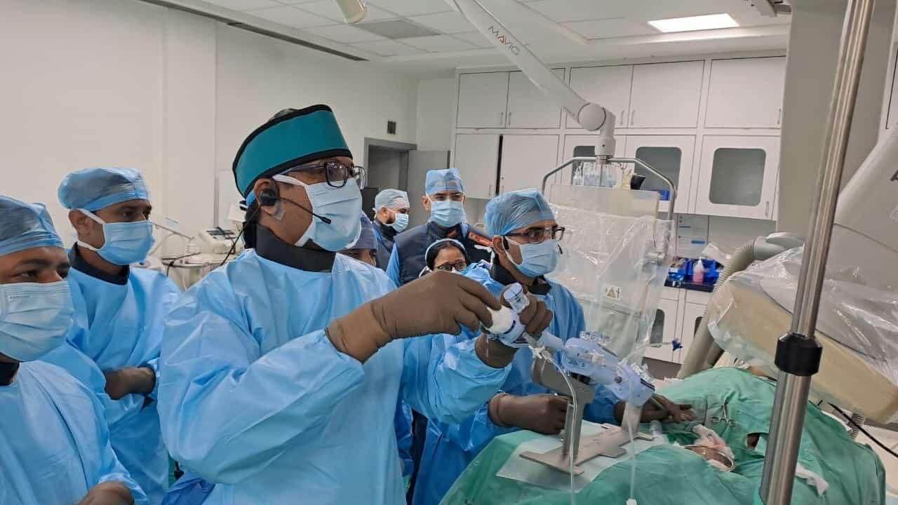
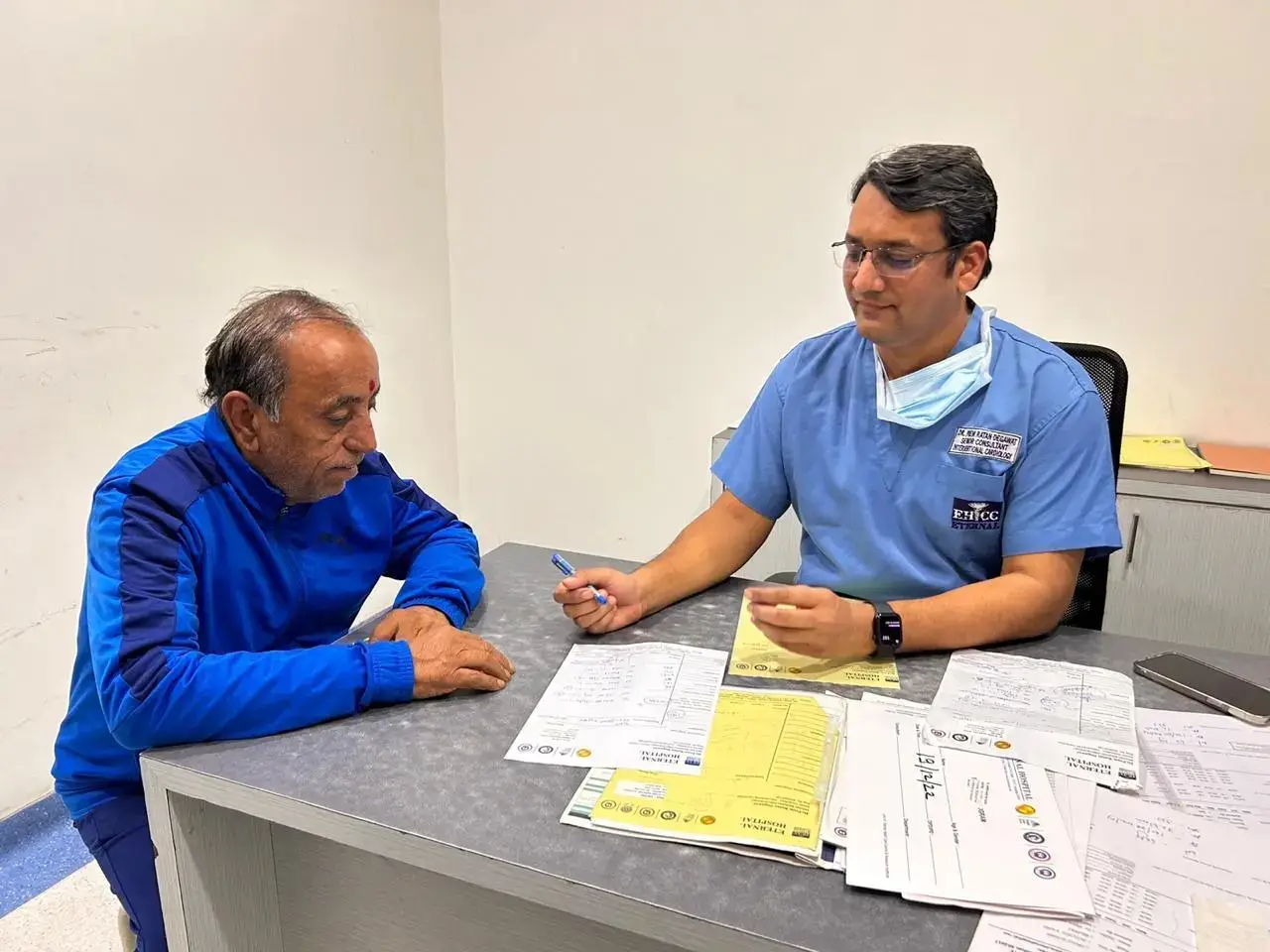



Dr. Prem Ratan Degawat is Associate Director of the TAVR & Structural Heart Disease Program and Director of the Mitral & Tricuspid Valve Program at Eternal Hospital, Jaipur.
He completed his DM in Cardiology at King George’s Medical University, Lucknow, and trained in advanced structural heart intervention at IRCCS Humanitas, Italy.
- Clinic: 6/384, In front of Railway Headquarter, Sector 6, Malviya Nagar, Jaipur, Rajasthan 302017
- Hospital for procedures: Eternal Hospital, 3A Jagatpura Road, Near Jawahar Circle, Jaipur 302017
- OPD: Monday to Saturday, 10:00 AM to 4:00 PM
- Contact: +91-8960594076
FAQs
Is stress echo better than TMT?
It gives different information by adding ultrasound pictures of the heart muscle. This can help when an exercise ECG is difficult to interpret or another question remains. But image quality, your ability to exercise and the reason for testing matter. Your doctor should choose the test that answers your particular question.
Does a stress echo show the percentage of blockage?
No. It looks for changes suggesting that blood supply becomes inadequate when the heart works harder. It does not directly measure each artery’s narrowing. If the result raises concern, your doctor may discuss artery imaging or other assessment, depending on your symptoms and the rest of your medical history.
Can I have a stress echo if I cannot walk on a treadmill?
Sometimes. Dobutamine can make the heart work harder while you lie down, allowing ultrasound pictures to be compared without treadmill exercise. It is not suitable for everyone. Tell the team why walking is difficult and about your medicines and heartbeat problems so it can choose an appropriate test.
Should I stop my beta blocker before the test?
Only if the clinician or testing centre specifically tells you to. Beta blockers slow the heartbeat and may affect the test, but stopping them can be unsafe in some people. Ask for written instructions for each medicine, including when to restart it, before changing your usual treatment schedule.
Can I ignore chest pain after a normal stress echo?
No. A normal result is useful but cannot exclude every cause of chest pain, especially if the test was limited. Persistent or changing symptoms need a review. Sudden severe pain, pain with marked breathlessness or sweating, or collapse needs emergency assessment regardless of an earlier normal test result.