Medically reviewed by Dr. Prem Ratan Degawat, MD, DM (Cardiology)
Senior Interventional Cardiologist · Associate Director, TAVR & Structural Heart Disease Program, Eternal Hospital, Jaipur
Last updated on April 4, 2026 · View LinkedIn profile
Your doctor has recommended an angiography. You heard the word and immediately felt uneasy.
That reaction is completely normal. Most patients arrive at this recommendation having never heard the term before. Others have heard it in alarming contexts, associated with surgery and serious diagnoses.
The reality is far less frightening than the reputation.
A coronary angiography is primarily a diagnostic test. Its job is to give your cardiologist a precise, real-time picture of the blood vessels supplying your heart. It does not automatically mean surgery. It does not mean the worst. In many cases, it simply gives the information needed to make the right decision.
Dr. Prem Ratan Degawat, interventional cardiologist at Eternal Hospital Jaipur, performs and interprets angiographies regularly. His approach begins with helping patients understand exactly what the test involves before they step into the procedure room.
What Coronary Angiography Actually Does
The heart is supplied by a network of arteries called coronary arteries. When one of these arteries narrows or becomes blocked, blood flow to a section of heart muscle is reduced.
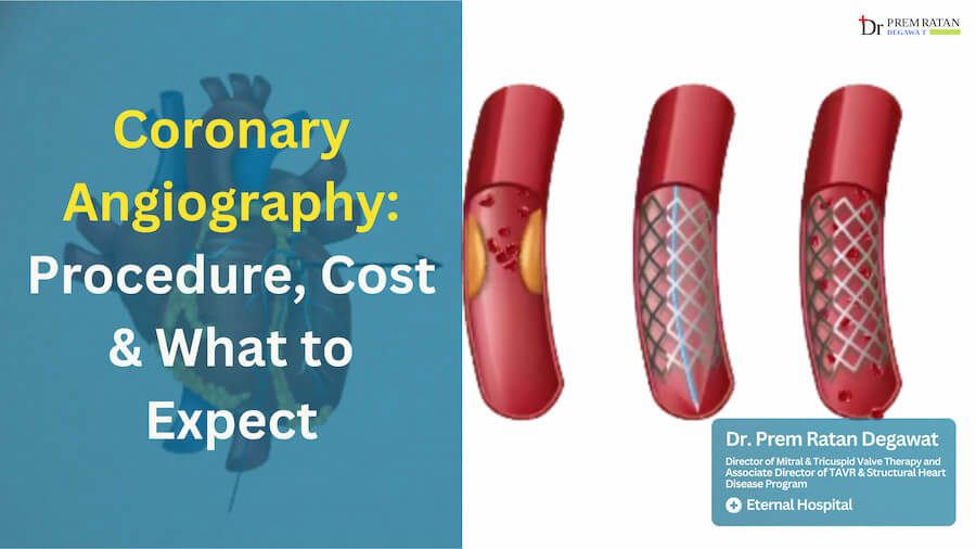
The problem is that these arteries are deep inside the body and invisible on a standard X-ray or ultrasound. Angiography solves this by injecting a special contrast dye directly into the coronary arteries through a thin tube called a catheter. Under live X-ray, this dye makes the arteries visible in real time on a screen.
Your cardiologist can see exactly where a narrowing starts, how severe it is, how long the blocked segment runs, and whether more than one artery is affected. No other test provides this level of detail.
This information determines everything that follows. Whether you need medication, a stent, or surgery depends entirely on what the angiography reveals.
When Does a Cardiologist Recommend Angiography?
Angiography is not a routine screening test. It is ordered when there is a specific clinical reason to look more closely at the coronary arteries.
Chest pain or tightness that appears during physical activity and disappears with rest is a classic indicator. This pattern, known as angina, suggests reduced blood flow to the heart under demand.
An abnormal stress test result is another common trigger. When the heart’s response to controlled exertion shows changes in the ECG, angiography is the logical next step to understand why.
Elevated cardiac enzymes in a blood test indicate that heart muscle cells have been damaged. This finding during a hospital admission typically leads to an urgent angiography.
If you have had a previous heart attack and your symptoms have changed or worsened, angiography helps assess whether existing blockages have progressed or new ones have developed.
Patients with multiple risk factors, such as diabetes, hypertension, high cholesterol, and a strong family history of heart disease, may be referred for angiography even before symptoms become severe, as a precautionary measure.
Preparing for Your Angiography: What to Do Before the Day
Good preparation reduces anxiety and helps the procedure go smoothly.
Several days before, your cardiologist will review your current medications. Blood thinners like warfarin or clopidogrel may need to be paused temporarily. Diabetic patients on certain oral medications may need to hold a dose or two around the procedure. Never stop any medication without explicit instruction from your doctor.
Blood tests, including kidney function, blood count, and clotting profile, are usually done in advance. The contrast dye used in angiography is filtered by the kidneys, so their baseline function matters.
On the day of the procedure, you must have nothing to eat or drink for at least four to six hours beforehand. Wear loose, comfortable clothing. Avoid jewellery and nail polish. Bring all previous cardiac reports, including any ECG, echo, or stress test results. Most importantly, bring a family member with you. You will not be in a position to drive yourself home.
A Step-by-Step Guide to What Happens During the Procedure
Understanding the sequence takes away much of the fear. Here is what actually happens.
You are changed into a hospital gown and shifted to the catheterization laboratory, or Cath Lab. This room contains specialised X-ray equipment and monitoring screens. It is temperature controlled and may feel cool.
A nurse checks your blood pressure, heart rate, and oxygen levels and attaches monitoring leads to your chest. An intravenous line is placed in your arm for medication delivery during the procedure.
The cardiologist cleans and numbs a small area at either your wrist (radial approach) or your upper thigh (femoral approach). Local anaesthetic is injected here. This is the only point of needle discomfort. You remain fully awake throughout the procedure. A mild sedative may be given to help you stay relaxed, but you are not put to sleep.
A thin, flexible catheter is guided through the artery to the opening of the coronary arteries. You will not feel this movement. The catheter travels through the blood vessel smoothly and causes no pain inside.
Once the catheter is positioned correctly, the contrast dye is injected in small amounts. You may feel a brief wave of warmth spread through your chest and body as the dye moves through. This sensation lasts only a few seconds and is completely harmless.
The cardiologist watches the dye travel through your coronary arteries on the X-ray screen in real time. Multiple images are taken from different angles to build a complete picture. The entire procedure takes between 30 and 45 minutes.
When imaging is complete, the catheter is removed. If the radial approach was used, a compression band is placed on your wrist. You are shifted to a recovery area to rest.
Reading Your Results: What the Three Possible Findings Mean
Patients are often most anxious about this part. Understanding the three main outcomes helps you process the results more clearly when your cardiologist explains them.
The first possibility is that the arteries are clean with no significant narrowing. This is good news, though your symptoms still need investigation through other tests. Non-cardiac causes of chest discomfort, such as acid reflux, muscle issues, or anxiety, can then be explored.
The second possibility is a moderate narrowing, typically in the range of 50 to 70 percent of the artery’s diameter. At this level, the blockage may not yet require a stent. Your cardiologist will likely recommend a combination of medications, dietary changes, and supervised exercise. Regular follow-up monitoring is important.
The third possibility is a severe narrowing of 70 percent or more, or a complete blockage. This level typically requires either coronary angioplasty with stenting or bypass surgery, depending on how many arteries are affected and where the blockages are located. In some cases, the cardiologist may be able to treat the blockage immediately during the same sitting, converting a diagnostic procedure into a therapeutic one.
Dr. Degawat takes time after every procedure to walk patients and their families through the images and explain what was found in clear, plain language. No decision about next steps is rushed.
Recovering After Angiography
Recovery from angiography is generally quick, especially with the radial (wrist) approach.
After the procedure, you rest in the observation area for four to six hours. Your blood pressure, heart rate, and the puncture site are monitored regularly. If the wrist approach was used, most patients are discharged the same evening. If the femoral (groin) approach was used, one night in hospital may be required.
For the first two days, avoid heavy lifting, strenuous activity, and prolonged standing. Drink plenty of water to help flush the contrast dye from your system. The puncture site may have minor bruising or slight tenderness, which is normal and resolves within a few days.
Contact the hospital immediately if you notice heavy bleeding or a rapidly growing bruise at the puncture site, numbness or coldness in the hand or leg on the same side, fever, chest pain, or difficulty breathing. These are rare but require prompt attention.
Angiography Cost in Jaipur: Government Schemes and Private Hospitals
The cost of angiography in Jaipur varies depending on the type of facility.
At government hospitals such as SMS Hospital and RUHS, angiography typically costs between Rs 3,000 and Rs 8,000. At private hospitals including Eternal Hospital, the range is approximately Rs 12,000 to Rs 25,000, depending on the complexity and the specific equipment used.
For patients enrolled in the Mukhyamantri Chiranjeevi Swasthya Bima Yojana, angiography is covered at empanelled centres. Eternal Hospital Jaipur is empanelled under this scheme. Carry your Jan Aadhaar card and inform the reception team at the time of admission. Ayushman Bharat PM-JAY cardholders are also eligible for coverage under applicable packages.
If you are unsure whether your procedure qualifies under a government scheme, the hospital’s insurance desk can verify your eligibility before the procedure date.
Is Angiography Safe? Understanding the Real Risk Picture
Coronary angiography has one of the strongest safety profiles of any invasive cardiac procedure. Millions of procedures are performed globally each year with very low complication rates.
Minor issues such as bruising at the puncture site, temporary discomfort, or a mild reaction to the contrast dye are relatively uncommon and resolve without intervention. Kidney function may be temporarily affected by the dye in patients with pre-existing kidney disease, which is why baseline kidney tests are done in advance and patients are advised to hydrate well afterward.
Serious complications are rare. A small number of patients, less than one in several hundred, may experience complications such as artery injury, allergic reaction to the dye, or cardiac arrhythmia during the procedure. These are managed immediately by the catheterization team. At Eternal Hospital Jaipur, full emergency cardiac support is available throughout every procedure.
Your cardiologist weighs the risk of the procedure against the risk of leaving a potentially serious cardiac condition undiagnosed. In the vast majority of cases, the information gained from angiography far outweighs the procedural risk.
About Dr. Prem Ratan Degawat


Dr. Prem Ratan Degawat is one of Jaipur’s most experienced interventional cardiologists, serving as Associate Director of the TAVR and Structural Heart Disease Program at Eternal Hospital, and as Director of the Mitral and Tricuspid Valve Program.
He completed his MBBS and MD from Sardar Patel Medical College, Bikaner, and his DM in Cardiology from King George’s Medical University, Lucknow. Advanced training in structural heart interventions was completed at IRCCS Humanitas Research Hospital in Italy.
With over 600 TAVI procedures and extensive experience in coronary intervention, Dr. Degawat brings both technical precision and a patient-centred approach to every case. He is known across Rajasthan for the time he takes to explain findings clearly, ensuring patients feel informed and confident at every stage of their care.
Consultation Details:
Hospital: Eternal Hospital, 3A Jagatpura Road, Near Jawahar Circle, Jaipur 302017
OPD Timings: Monday to Saturday, 10:00 AM to 4:00 PM
Clinic: 6/384, infront of railway headquarter, Sector 6, Malviya Nagar, Jaipur, Rajasthan 302017
Contact: +91-8960594076
FAQs
Is Coronary Angiography Painful?
Only the local anaesthetic injection at the puncture site causes brief discomfort. The catheter movement inside the artery is not felt at all. Most patients find the procedure far more comfortable than they expected.
How Much Does Angiography Cost in Jaipur?
Private hospitals charge between Rs 12,000 and Rs 25,000. Government hospitals like SMS and RUHS range from Rs 3,000 to Rs 8,000. Chiranjeevi Yojana and Ayushman Bharat cardholders can access cashless coverage at Eternal Hospital Jaipur.
Will I Be Unconscious During the Procedure?
No. You stay fully awake. Local anaesthetic is applied only at the puncture site. A mild sedative may be given to keep you relaxed, but general anaesthesia is not used.
How Long Does Recovery Take After Angiography?
With the wrist approach, most patients go home the same day. Avoid heavy activity for two to three days. Most people return to their normal routine within three to four days.
What Is the Difference Between Angiography and Angioplasty?
Angiography is a diagnostic test that identifies blockages. Angioplasty is the treatment that opens those blockages using a balloon and stent. In some cases, both are performed in the same sitting.
Is Angiography Covered Under Chiranjeevi Yojana?
Yes. It is included under the Chiranjeevi Yojana package at empanelled hospitals. Carry your Jan Aadhaar card and inform the admissions desk before your procedure date.
What Should I Avoid Eating Before the Test?
Do not eat or drink anything for four to six hours before the procedure. Follow your doctor’s specific instructions for diabetes or blood pressure medications before fasting.
Can Angiography Be Done on the Same Day as Consultation?
For planned cases, no. Pre-procedure blood tests, medication review, and fasting are required beforehand. In emergency situations, same-day angiography is carried out immediately.
The information in this article is for general awareness only. It is not a substitute for professional medical advice, diagnosis, or treatment.